
The liver removes substances from our blood, but parts of this system become less efficient as we age.
Scientists have now restored some of this lost function in older rats using an experimental drug, cutting blood levels of a protein fragment linked to Alzheimer’s disease by half.
The treatment also reduced cholesterol. After this second discovery, the researchers discovered that the drug restored communication between neighboring liver cells.
The study, led by researchers at the University of Southern California, focused on protein fragments that can build up as plaques in the brains of people with Alzheimer’s disease. Called beta-amyloid, these fragments also circulate in the blood, where the liver helps eliminate them.
Restoring the clearance capacity of the aging liver could therefore reduce circulating beta-amyloid, thereby addressing a consequence of aging that could influence disease risk.
“Age is the greatest risk factor for late-onset Alzheimer’s disease,” Sam de Leve, first author and liver researcher at USC, told ScienceAlert.
To investigate, the team looked at how the substances reach the cells responsible for processing them. Much of the beta-amyloid in the blood travels attached to fat-carrying particles. The large particles must pass through microscopic openings in cells lining the liver’s small blood vessels to reach its main working cells, called hepatocytes.
As the cells lining the vessels age, these openings become smaller and smaller. The researchers suspected that reopening them would help hepatocytes collect beta-amyloid from the blood.
They gave two-year-old rats an experimental drug called cinaciguat daily for two weeks, comparing them to older, untreated rats and four-month-old animals.
The treatment restored the openings. Blood levels of amyloid beta 40, a form of the protein fragment, fell by half, returning to youthful levels.
LDL, the cholesterol-carrying particles commonly known as “bad cholesterol,” also decreased. However, LDL particles are small enough to pass through the vessel’s interior lining even when its openings are lost. Reopening these passages could not explain this result.
The team tested whether the drug also affected communication between cells. They collected a fluid in which cells lining the vessels of young or old rats had grown overnight, then added it to the hepatocytes of young rats.
Fluid from young cells promoted the absorption of LDL. Fluid from old cells provided no more support than regular culture medium. However, when older donor rats were given cinaciguat, fluid from their cells again promoted absorption.
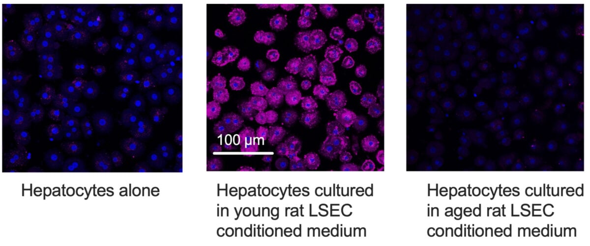
The treatment appears to have restored a message helping hepatocytes maintain their specialized function. De Leve compared this to a scene in The Lion King: After the death of his father, Simba flees his future as king, until his father, Mufasa, appears to him in spirit.
“It’s like Mufasa telling Simba to ‘Remember who you are’ in The Lion King” said de Levé.
In the liver, this booster came via a growth factor, a protein that influences cellular behavior, called HB-EGF. Older cells lining the vessels still produced its precursor, but did not release detectable amounts of soluble signal. Cinacigua has restored this version.
Addition of HB-EGF directly to hepatocytes enhanced their uptake of LDL and beta-amyloid. Blocking it removed the benefits provided by the fluid from young cells.
Hepatocytes offered another clue. Two proteins that capture these substances were present in similar total amounts in older cells, but they were found in lower amounts on the cell surface, where they act. The treatment restored their surface levels.
The results support the role of HB-EGF in this change, although the researchers did not directly establish this link.
De Leve explained that restoring the cells lining the vessels both reopened the passages for large molecules and renewed their communication with hepatocytes.
“In youth, the hepatocyte is a specialist in maintaining appropriate circulating levels of LDL cholesterol and beta-amyloid, but with aging, the hepatocyte becomes less efficient at removing these substances,” de Leve said.
It remains unclear whether improving liver clearance could protect the brain. Previous research in mice has linked amyloid produced in the liver to changes in the brain.
This study examined healthy aging, using three to six independent biological samples per condition in different experiments. The treatment has not been shown to reduce brain plaques, improve memory or prevent Alzheimer’s disease.
The researchers say this approach should next be tested in an animal model of Alzheimer’s disease. Developing a human treatment would also require further research into the effectiveness and safety of related drugs.
The research was published in Journal of Biological Chemistry.
This article was fact-checked by Rebecca Dyer and edited by Rebecca Dyer. Although we are proud of our process, we are only human. If you spot an error, please let us know.
Gn Health